
The Precision Imaging Vanguard: How Advanced Radiodiagnosis and Sonoelastography Are Transforming Early Detection in Indian Oncology and Neurology
5 min read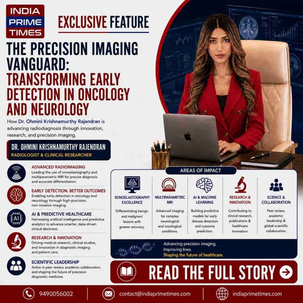
Modern clinical medicine is undergoing a profound structural shift toward non-invasive diagnostics. For decades, the definitive diagnosis of complex internal pathologies-ranging from suspicious cervical lymphadenopathy to intracranial brain lesions-relied heavily on invasive tissue biopsies or exploratory surgical procedures. While effective, these invasive interventions carry notable patient discomfort, procedural risk, and significant clinical delays. Today, rapid technological advancements in medical imaging are rewriting that paradigm. The contemporary field of Radiodiagnosis is no longer confined to merely visualizing static anatomical structures; it has evolved into a dynamic science of functional, multiparametric tissue characterization.
Across India’s leading healthcare hubs, diagnostic specialists are deploying advanced imaging modalities that can differentiate between benign inflammation, infectious pathologies, and aggressive malignancies at an unprecedented cellular and mechanical level. To investigate how these high-precision imaging protocols are being operationalized within high-volume clinical settings, the India Prime Times editorial team recently conducted extensive reporting across key medical and oncology corridors in Bengaluru.
During our field reporting across tertiary teaching institutions and dedicated cancer care centers, our team observed the critical role that real-time radiological interpretation plays in shaping treatment trajectories. In high-stakes emergency and oncology multidisciplinary tumor boards, surgical oncologists, neurologists, and physicians rely entirely on the diagnostic precision of the radiologist before making a single surgical incision.
This vital clinical dynamic led our editorial desk to an in-depth interaction with Dr. Ohmini Krishnamurthy Rajendran, an accomplished Radiologist and Clinical Researcher. Practicing as a Senior Resident in Radiodiagnosis at the Kempegowda Institute of Medical Sciences (KIMS) and Research Hospital, and serving as a Consultant Radiologist at Janasnehi Diagnostics, BRINDHAVVAN AREION HOSPITAL, and Arion Radiotherapy and Oncology Centre, Dr. Ohmini represents a new wave of diagnostic specialists bridging academic research with demanding frontline clinical practice.
Over a comprehensive conversation, the India Prime Times team explored her research into tissue elasticity, advanced neuro-imaging, and the evolving mandate for non-invasive diagnostic precision.
Redefining Lymph Node Characterization Through Sonoelastography
A persistent diagnostic challenge in clinical oncology is the assessment of suspicious lymph nodes. Lymph nodes frequently enlarge in response to routine viral infections, tuberculosis, or malignant metastatic spread from distant primary tumors. Distinguishing between reactive lymphadenopathy and metastatic infiltration using conventional B-mode ultrasound alone can often be ambiguous, frequently leading to unnecessary Fine Needle Aspiration Cytology (FNAC) or surgical excision. During our discussion, Dr. Ohmini highlighted her scholarly research focused on “Sonoelastography in Characterization of Suspicious Lymph Nodes and Correlating with Histopathology.” “Conventional ultrasound primarily assesses morphological parameters such as shape, size, and vascularity,” Dr. Ohmini explained to the India Prime Times team. “However, malignant transformations fundamentally alter the physical stiffness of tissue due to dense cellular proliferation and desmoplasia. Sonoelastography allows us to map tissue elasticity non-invasive in real time. When we correlate elasticity scores with gold-standard histopathological findings, we achieve a much higher level of diagnostic accuracy, enabling clinicians to identify malignancy earlier while sparing benign cases from invasive biopsies.” Her research underscores how quantitative elasticity assessment provides an objective, non-invasive biomarker, directly strengthening oncological staging, surgical planning, and early therapeutic decision-making.
Navigating the Neurological Labyrinth: Multiparametric MRI for Ring-Enhancing Lesions
Beyond superficial tissue assessment, our conversation delved into the complex domain of neuroradiology. In tropical and developing regions like South Asia, intracranial ring-enhancing lesions (RELs) represent one of the most difficult diagnostic puzzles confronting radiologists and neurosurgeons. A ring-enhancing lesion on a brain scan can represent an array of vastly divergent conditions:
Infectious and Parasitic Etiologies: Such as neurocysticercosis, tuberculomas, or cerebral abscesses. Primary Malignancies: High-grade gliomas and glioblastomas. Metastatic Carcinomas: Secondary lesions spread from lung, breast, or gastrointestinal cancers. Demyelinating and Inflammatory Disorders: Such as tumefactive multiple sclerosis. Misdiagnosing an infectious lesion as a malignant brain tumor can subject a patient to unnecessary craniotomy or radiation, while mistaking a glioblastoma for an abscess delays critical oncological therapy.
Dr. Ohmini addressed this clinical dilemma in her extensive postgraduate thesis, titled “Multiparametric MRI Approach to Ring Enhancing Lesions.” Completed during her MD in Radiodiagnosis from Kempegowda Institute of Medical Sciences (2025)-following her foundational MBBS from MVJ Medical College, Bangalore-her work evaluates how combining advanced MRI sequences provides definitive differential clarity. “Relying on conventional T1-weighted contrast enhancement alone is insufficient for complex intracranial lesions,” Dr. Ohmini noted. “By deploying a multiparametric MRI protocol-integrating Diffusion-Weighted Imaging (DWI), Magnetic Resonance Spectroscopy (MRS), and Perfusion-Weighted Imaging (PWI)-we analyze cellular density, microscopic water diffusion, and metabolite ratios within the lesion wall and core. This multi-sequence assessment allows us to confidently differentiate a pyogenic abscess from a necrotic high-grade glioma without opening the skull.”
The Multi-Disciplinary Radiologist in Modern Healthcare
What stood out during our team’s observations at BRINDHAVVAN AREION HOSPITAL and Arion Radiotherapy and Oncology Centre is how modern radiology has moved out of isolated dark rooms and into the center of multidisciplinary patient care. Dr. Ohmini’s day-to-day practice encompasses a broad diagnostic spectrum, including Computed Tomography (CT), high-resolution Magnetic Resonance Imaging (MRI), emergency trauma radiography, and ultrasound-guided interventional procedures such as FNACs. Her reporting directly informs radiotherapy planning target volumes, surgical resectability criteria, and real-time emergency room stabilizations. “Radiology is fundamentally an investigative science of clinical correlation,” she emphasized during our interview. “High diagnostic accuracy is not just about identifying an abnormality on a screen; it requires correlating that finding with the patient’s clinical history, biochemistry, and pathological markers to deliver a report that gives treating physicians an actionable roadmap.”
Extending Radiology Through Research, Artificial Intelligence, and Medical Innovation
Beyond her clinical practice, Dr. Ohmini’s work reflects a broader focus on integrating advanced medical imaging with emerging technologies, artificial intelligence, and data-driven approaches to precision healthcare. Her research interests extend across diagnostic imaging, oncology, neurological imaging, predictive healthcare, and the development of innovative approaches aimed at improving early disease detection and clinical decision-making. This multidisciplinary approach reflects her vision of radiology evolving from conventional image interpretation toward increasingly quantitative, predictive, and technology-enabled diagnostic medicine. Her academic contributions include peer-reviewed scientific publications, research in advanced diagnostic imaging, and participation in the peer-review process for scientific journals. Her work has also extended into medical innovation and intellectual property, with multiple healthcare-related utility patents developed around diagnostic and clinical applications. Her growing academic portfolio includes 18 peer-reviewed publications, more than 850 citations on ResearchGate and more than 745 citations on Google Scholar, approximately 90 scientific peer reviews, and 10 Indian utility patents, reflecting sustained engagement in medical research, scientific evaluation, and healthcare innovation. Through this combination of clinical practice, research, scientific peer review, and innovation, Dr. Ohmini is contributing to the evolution of precision diagnostic medicine, with a focus on advancing accurate, non-invasive, and technology-enabled healthcare in India and internationally.
The Future of Diagnostic Medicine
As our conversation with Dr. Ohmini Krishnamurthy Rajendran concluded, the overarching trajectory of India’s diagnostic landscape came into sharp focus. The future of healthcare delivery will be determined by how swiftly and accurately diseases can be detected before structural damage occurs. For hospital administrators, oncologists, and clinicians navigating the increasing complexities of chronic disease and cancer care, the methodological rigor demonstrated by specialists like Dr. Ohmini Krishnamurthy Rajendran provides a vital benchmark. By uniting evidence-based clinical acumen, advanced non-invasive imaging techniques like sonoelastography, and comprehensive multiparametric MRI protocols, modern radiologists are ensuring that Indian diagnostic medicine remains precise, patient-centric, and globally competitive.